
- Application Article
- Download
Hooke College

- About Us
- Team
- R & D and production
- Join Us
- Contact Us
- Qualification honor


The explosive growth in biomedical research using live-cell imaging technology has been driven by a series of events, including dramatic advances in confocal microscopy instruments, the introduction of novel ultra-sensitive detectors, and continued improvements in the performance of genetically encoded fluorescent proteins. The acquisition of images of local fluorophores in living cells on millisecond timescales, revealing complex biodynamics, presents many new challenges that are far more complex than traditional problems. To ensure high image acquisition speeds for fluorescent proteins and synthetic dyes in living cells, with reasonable contrast and minimal photobleaching, the microscope must be able to quickly scan the field of view and record data using detectors with high quantum efficiency. Laser scanning confocal microscopy focuses a single beam on the sample plane to sequentially point scan the region of interest and spatially filters the emitted light through a single pinhole. This approach limits the speed of image acquisition due to the need for extremely precise control of the galvanometer mirror used to scan the beam on the raster sample and the limited number of photons emitted by the sample for pixel dwell time. For general imaging scenes, confocal microscopes scan at 1 ms per pixel, which means that each image is acquired at a rate of between one-half and two seconds, depending on the size. As a result, most laser-scanning confocal microscopes are not sufficient to capture the dynamic events at the millisecond scale that are often crucial for unraveling the complex molecular processes occurring in living cells. Although the scanning speed limitation can be overcome by employing a resonant scanning scheme, this high-speed acquisition has ultra-short pixel dwell times and therefore insufficient SNR.
Many of the speed limitations associated with point-scanning confocal microscopy can be overcome by imaging the sample using multiple excitation beams operating in parallel, that is, turntable confocal microscopy. At the same time, the improvement of light utilization makes the softer LED light source can replace the laser light source, which greatly reduces the light damage during the observation process. Turntable confocal microscopy is emerging as a powerful tool for rapid spatial and temporal imaging of living cells. Although the technique was originally introduced more than 40 years ago, recent improvements in microscope optical design and camera technology have significantly expanded the versatility and potential of this approach.
The main purpose of developing confocal imaging sharpness enhancement algorithm in this project is to correct the fuzzy and unclear problems in confocal 3D imaging, and present important image details, so as to realize clear and accurate imaging of 3D structure of biological samples, and observe the dynamic changes inside living cells.
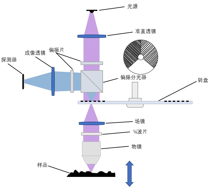
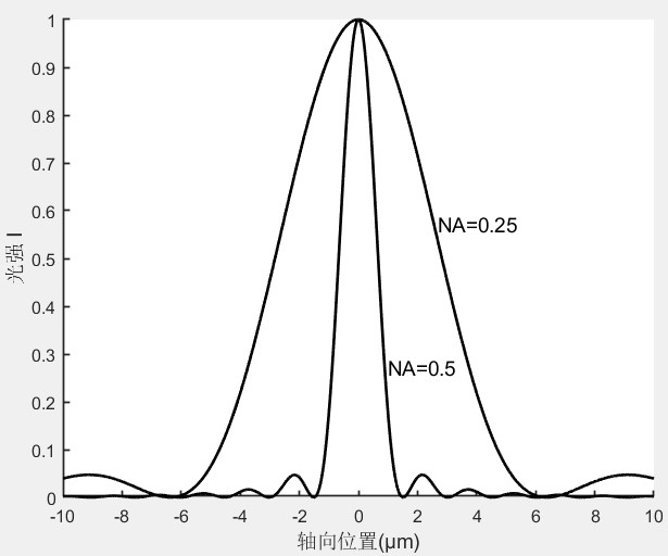
The turntable confocal microscope (Figure 1) uses a mask plate to limit the range of illumination light and prevent defocus light from participating in the imaging, thus greatly improving the axial resolution (with a certain increase in the lateral resolution), which enables the confocal microscope to have a narrow axial response and scan the sample layer by layer. The axial response curve of Gaussian distribution as shown in FIG. 2 can be obtained at each point on the surface, and the spatial height of the point can be obtained by finding the maximum value through Gaussian fitting. Then, the three-dimensional morphology of the sample surface can be reconstructed, and surface defect detection, roughness detection and physical property analysis can be carried out. At present, the lateral resolution of relevant instruments on the market can reach 120-140μm, and the axial resolution is 1-0.1nm. Compared with the point scanning method, the rotary disk scanning method greatly improves the scanning speed with a small sacrifice of resolution.

FIG. 1 Schematic diagram of confocal turntable

FIG. 2 Confocal axial response curve
In microscopic image analysis, the application of 3D imaging system is very important. Since the morphology of cells can not be observed by the naked eye, the sequence images can only be obtained by magnification of the microscope, and then the 3D reconstruction technology of computer is used to reconstruct and display the 2D sequence images, so that the 3D images of the cell structure and the images at different depth levels can get the real 3D morphology display.

+86-431-81077008
+86-571-86972756

Building 3, Photoelectric Information Industrial Park, No.7691 Ziyou Road, Changchun, Jilin, P.R.C
F2006, 2nd Floor,South Building, No. 368 Liuhe Road, Binjiang District, Hangzhou, Zhejiang,P.R.C

sales@hooke-instruments.com

COPYRIGHT©2022 HOOKE INSTRUMENTS LTD.ALL RIGHTS RESERVED 吉ICP备18001354号-1